| --- |
| pretty_name: "REYIA: The Largest Multi-Source Retinal Artery/Vein Segmentation Dataset" |
| license: other |
| license_name: apache-2.0-plus-third-party |
| license_link: LICENSE |
| task_categories: |
| - image-segmentation |
| tags: |
| - retinal-fundus |
| - artery-vein-segmentation |
| - medical-imaging |
| - ophthalmology |
| size_categories: |
| - n<1K |
| viewer: false |
| extra_gated_heading: "Request access to REYIA" |
| extra_gated_description: >- |
| REYIA bundles ENRICH, a dataset of retinal fundus images collected under |
| KU Leuven Hospital Helsinki Committee approval (S66963). Access requests |
| are logged to keep a record of who is using the data and for what |
| purpose, per that approval's terms. |
| extra_gated_prompt: >- |
| By requesting access you agree to use ENRICH (images and annotations) for |
| non-commercial purposes only, per its KU Leuven data-sharing protocol. |
| REYIA's own annotations and code are Apache 2.0, and source images from |
| the other subsets follow their own original licenses (see below). You |
| agree to cite the REYIA paper, the Lirot.ai paper (the platform used to |
| produce REYIA's annotations), and each original subset's own citation in |
| any resulting publication. |
| extra_gated_fields: |
| Full name: text |
| Affiliation (institution/lab): text |
| Intended research use: text |
| I agree to the license terms above (ENRICH is non-commercial; see the License section for the rest) and to cite REYIA, the annotation platform, and the original subsets: checkbox |
| extra_gated_button_content: "Agree and request access" |
| --- |
| |
| # REYIA: The Largest Multi-Source Retinal Artery/Vein Segmentation Dataset |
|
|
| <div align="center"> |
|
|
| Jonathan Fhima • Jan Van Eijgen • Lennert Beeckmans • Thomas Jacob • Moti Freiman |
| <br> |
| Luis Filipe Nakayama • Ingeborg Stalmans • Chaim Baskin • Joachim A. Behar |
|
|
| </div> |
|
|
| [](https://arxiv.org/abs/2503.01190) |
| [](https://github.com/aim-lab/RLAD) |
| [](https://huggingface.co/JonathanFhima/RLAD-SwinV2-Large) |
| [](https://huggingface.co/datasets/JonathanFhima/REYIA/blob/main/LICENSE) |
|
|
|  |
|
|
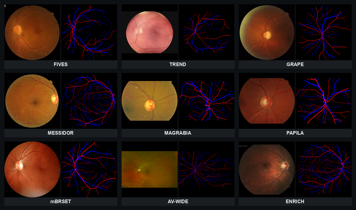
| **REYIA** is a curated set of 589 retinal fundus images annotated with AV |
| (artery/vein) blood vessel segmentations using the open-access |
| [Lirot.ai](https://www.aimlab-technion.com/lirot-ai) software. To enhance |
| diversity, REYIA includes manually segmented images from nine sources: |
| [FIVES](https://www.nature.com/articles/s41597-022-01564-3), |
| [TREND](https://pmc.ncbi.nlm.nih.gov/articles/PMC8301647/), |
| [GRAPE](https://www.nature.com/articles/s41597-023-02424-4), |
| [MESSIDOR](https://www.adcis.net/en/third-party/messidor2/), |
| [MAGRABIA](https://deepblue.lib.umich.edu/data/concern/data_sets/3b591905z), |
| [PAPILA](https://figshare.com/articles/dataset/PAPILA/14798004), |
| [mBRSET](https://physionet.org/content/mbrset/1.0/), |
| [AV-WIDE](https://people.duke.edu/~sf59/Estrada_TMI_2015_dataset.htm), and |
| ENRICH, a new dataset of 111 images collected for this study. AV-WIDE, |
| which originally shipped only skeletonized vessel traces, was reannotated |
| here with complete AV segmentations. |
|
|
| This repository ships the annotations for all 589 images, plus the source |
| images themselves wherever their license allows redistribution. For the |
| rest, it ships a script that reconstructs the exact image REYIA annotated |
| from your own raw download. |
|
|
| ## License |
|
|
| **REYIA's own annotations and the code in `scripts/` are licensed under |
| Apache 2.0** (see |
| [`LICENSE`](https://huggingface.co/datasets/JonathanFhima/REYIA/blob/main/LICENSE)), |
| with one exception: **ENRICH — both the images and their annotations — is |
| non-commercial use only**, per the KU Leuven Hospital data-sharing protocol |
| under which it was collected (Helsinki approval S66963). Source images |
| from the other eight datasets keep their own original licenses: |
|
|
| | Subset | Bundled here | License | |
| |---|---|---| |
| | FIVES | Yes | CC BY 4.0 | |
| | GRAPE | Yes | CC0 1.0 | |
| | TREND | Yes | CC BY 4.0 | |
| | ENRICH | Yes | Non-commercial use only — KU Leuven data-sharing protocol (own data) | |
| | PAPILA | No | figshare's license field reads GPL 3.0+ (most likely a depositor error — GPL is a software license, not meant for a dataset — but never corrected upstream). Images not bundled here | |
| | MAGRABIA | No | Deep Blue Data tags the full RIGA release CC BY-NC 4.0; no subset-specific scope is published for Magrabia alone. Images not bundled here | |
| | MESSIDOR | No | Explicit no-redistribution terms | |
| | mBRSET | No | PhysioNet Data Use Agreement, credentialed access only | |
| | AV-WIDE | No | Explicit no-redistribution terms | |
|
|
| Annotations for every subset other than ENRICH are released here under |
| Apache 2.0 regardless of the source image's own license. ENRICH's |
| annotations follow ENRICH's own non-commercial restriction above. |
|
|
| ## Download & build |
|
|
| ```bash |
| python3 -m venv reyia_env && source reyia_env/bin/activate |
| pip install huggingface_hub |
| huggingface-cli download JonathanFhima/REYIA --repo-type dataset --local-dir REYIA |
| cd REYIA |
| ``` |
|
|
| FIVES, GRAPE, TREND, and ENRICH are already bundled — use `images/<subset>/` |
| and `annotations/<subset>/` directly. |
|
|
| For the other five, download each one and extract it — as is, no need to |
| pick files out — into `raw_downloads/<subset>/` (you don't need all five; |
| subfolders inside are fine, everything is found recursively): |
|
|
| | Subset | Download | Extract into | What's in there | |
| |---|---|---|---| |
| | PAPILA | https://figshare.com/articles/dataset/PAPILA/14798004 | `raw_downloads/papila/` | the `.jpg` fundus photos | |
| | MAGRABIA | https://deepblue.lib.umich.edu/data/concern/data_sets/3b591905z | `raw_downloads/magrabia/` | keep the `MagrabiFemale/` and `MagrabiaMale/` folders as-is — their names are how the script tells images apart | |
| | MESSIDOR | https://www.adcis.net/en/third-party/messidor2/ (registration required) | `raw_downloads/messidor/` | just the `Base11` package (one of 9 zips) — the `.tif` files | |
| | mBRSET | https://physionet.org/content/mbrset/1.0/ (credentialed access) | `raw_downloads/mbrset/` | the `images/` folder from the download | |
| | AV-WIDE | https://people.duke.edu/~sf59/Estrada_TMI_2015_dataset.htm | `raw_downloads/av-wide/` | the `AV-WIDE/` folder of `.mat` files | |
|
|
| ```bash |
| pip install -r scripts/build/requirements.txt |
| bash scripts/build/build_reyia.sh |
| ``` |
|
|
| This writes each reconstructed image into `images/<subset>/`, matched to |
| its `reyia_id`. |
|
|
| ## Ethical Consent |
|
|
| **ENRICH.** Approved by the Helsinki Committee at KU Leuven Hospital |
| (approval S66963); informed consent waived, all identifiable patient |
| information removed. |
|
|
| **GRAPE.** Registered clinical study (ClinicalTrials.gov NCT04718532), |
| approved by Ethics Committee of ZJU-2 (No Y2020-1027). |
| https://www.nature.com/articles/s41597-023-02424-4 |
|
|
| **MESSIDOR.** Free for research/educational use; redistribution and |
| unauthorized commercial use prohibited; publications must acknowledge |
| LaTIM laboratory and Messidor program partners. |
| https://www.adcis.net/en/third-party/messidor2/ |
|
|
| **PAPILA.** Collected at Hospital General Universitario Reina Sofía |
| (Murcia, Spain), 2018–2020, under hospital Ethics Committee approval with |
| patient informed consent. |
| https://www.nature.com/articles/s41597-022-01388-1 |
|
|
| **MAGRABIA.** Ethical approval obtained for the de-identified dataset from |
| University of Waterloo, Canada. |
| https://www.spiedigitallibrary.org/conference-proceedings-of-spie/10579/2293584/Retinal-fundus-images-for-glaucoma-analysis-the-RIGA-dataset/10.1117/12.2293584.full |
|
|
| **FIVES.** Approved by the Medical Ethics Committee of the Second |
| Affiliated Hospital of Zhejiang University; written informed consent from |
| every participant; registered on ClinicalTrials.gov (NCT04718532). |
| https://www.nature.com/articles/s41597-022-01564-3 |
|
|
| **AV-WIDE.** Acquired at Duke University Medical Center under Duke IRB |
| protocols Pro00015512 and Pro00056311. |
| https://people.duke.edu/~sf59/estradaTMI15.pdf |
|
|
| **TREND.** Approved by the Ethical Committee of the Faculty of Medicine, |
| University of Montenegro (Protocol No. 2487/4 and 3824/4 / 03/01-11417/1). |
| https://pmc.ncbi.nlm.nih.gov/articles/PMC8301647/ |
|
|
| **mBRSET.** Approved by the Institutional Review Board of IPTAN (protocol |
| CAAE 64219922.3.0000.9667); all identifiable patient information removed; |
| written consent obtained for image capture and open publication. |
| https://www.nature.com/articles/s41597-025-04627-3 |
|
|
| ## Benchmark reference |
|
|
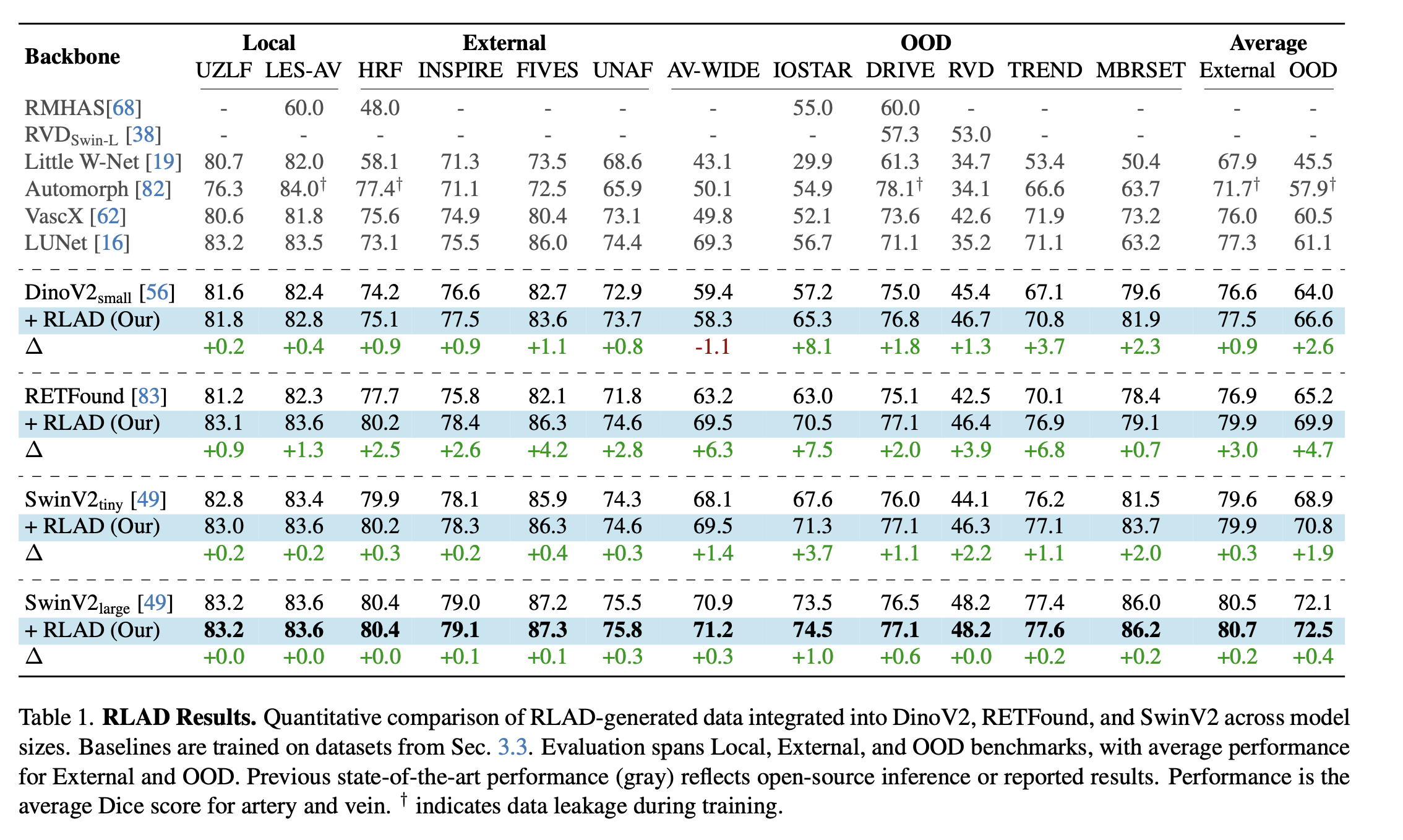
| If you train a model on REYIA (or on data augmented with it), these are |
| our own reference numbers — artery/vein Dice score, averaged — across the |
| same local, external, and out-of-distribution (OOD) test sets, so you can |
| compare directly instead of re-running every baseline yourself. `∆` rows |
| show the improvement RLAD adds on top of each backbone. `†` marks results |
| with data leakage during training. We recommend comparing against |
| [RLAD-SwinV2-Large](https://huggingface.co/JonathanFhima/RLAD-SwinV2-Large), |
| the strongest model in this table and the one we release weights for. |
|
|
|  |
|
|
| `scripts/eval/dice_score.py` is the exact scorer used to produce these |
| numbers — artery is the mask's red channel, vein is the blue channel, both |
| nonzero after thresholding, so magenta crossing pixels count toward both. |
|
|
| ```bash |
| pip install -r scripts/eval/requirements.txt |
| python3 scripts/eval/eval_example.py --mask annotations/<subset>/reyia_XXXX.png |
| ``` |
|
|
| This scores a real REYIA mask against itself (prints 1.0 across the board, |
| as a sanity check). |
|
|
| ## Citation |
|
|
| If you use REYIA, please cite the paper it was introduced in: |
|
|
| > Fhima, J., Van Eijgen, J., Beeckmans, L., Jacob, T., Freiman, M., |
| > Nakayama, L.F., Stalmans, I., Baskin, C., Behar, J.A. *Enhancing Retinal |
| > Vessel Segmentation Generalization via Layout-Aware Generative |
| > Modelling.* arXiv:2503.01190, 2025. https://arxiv.org/abs/2503.01190 |
|
|
| REYIA's annotations were produced with [Lirot.ai](https://www.aimlab-technion.com/lirot-ai). |
| Please also cite the paper that introduced it: |
|
|
| > Fhima, J., Van Eijgen, J., Freiman, M., Stalmans, I., Behar, J.A. *Lirot.ai: |
| > A Novel Platform for Crowd-Sourcing Retinal Image Segmentations.* 2022 |
| > Computing in Cardiology (CinC), vol. 498, pp. 1-4, IEEE, 2022. |
| > arXiv:2208.10100. https://arxiv.org/abs/2208.10100 |
|
|
| Code (RLAD): https://github.com/aim-lab/RLAD |
|
|
| Please also cite the original dataset(s) any images you use come from, per |
| their own citation requirements (see the Ethical Consent section above). |
|
|

